
In this post, you can view and download all the important Histology slides in the 1st year of MBBS.
It includes both general and systemic slides. Along with the pictures of the histological slides, two identification points have also been mentioned for your understanding and convenience.
You can bookmark this page for a quicker revisit. 💪
I have also included a PDF that you can download, at the end of this blog. ❤️
There are dozens of Histology Textbooks out there, making it difficult for medical students to decide which ones to purchase. To make it easier for you, I’ve handpicked BEST and MOST RECOMMENDED textbooks:
You might also be interested to check out NMC’s recommended list of books for MBBS 1st Year.
Cartilage
Hyaline cartilage

Points of identification:
- Cell nests of chondrocytes present.
- Territorial and interterritorial matrix present.
- Perichondrium present.
Yellow elastic cartilage

Points of identification:
- Large singly arranged chondrocytes in lacunae.
- Perichondrium present.
White fibrocartilage

Points of identification:
- Chondrocytes of similar size present between collagen bundles.
- Perichondrium absent.
Compact Bone
Transverse section of bone
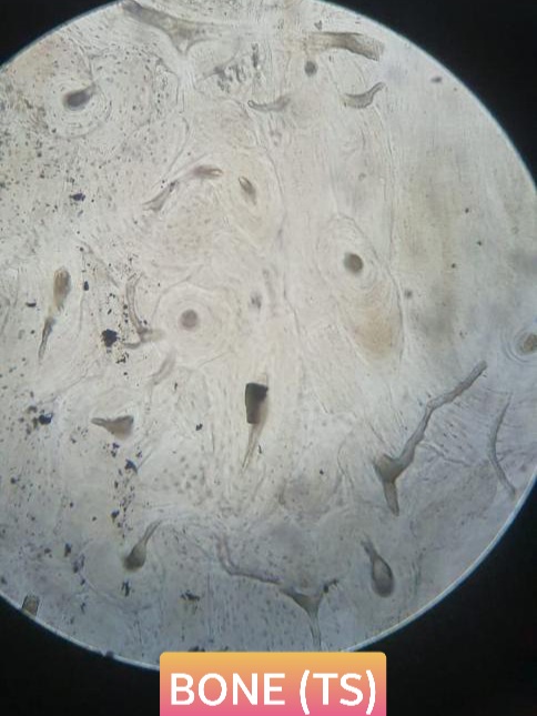
Points of identification:
- Presence of osteocytes in lacunae.
- Haversian system present with concentric lamellae.
Longitudinal section of bone
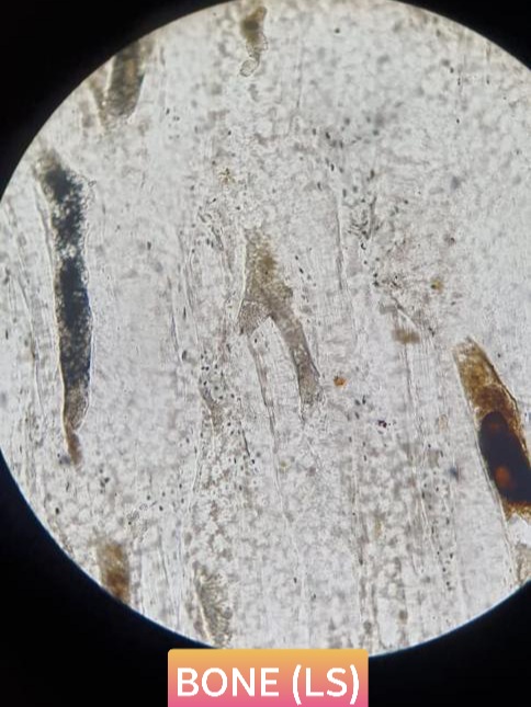
Points of identification:
- Osteocytes present.
- Longitudinal section of the haversian system and Volkmann’s canal seen.
Muscular Tissue
Skeletal muscle

Points of identification:
- Cylindrical muscle fibers with prominent striations.
- Presence of peripherally arranged flattened multinuclei.
Cardiac muscle

Points of identification:
- Branching fibers with striations present.
- Intercalated discs and centrally placed nucleus present.
Nervous Tissue
Peripheral nerve (transverse section)

Points of identification:
- Nerve fibres arranged in fasiculi.
- Endoneural, perineual and epineural connective tissues seen.
Optic nerve (transverse section)
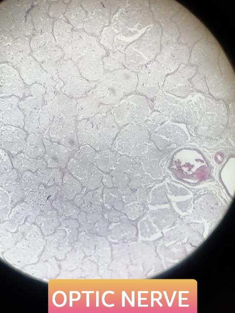
Points of identification:
- Presence of 3 layers of meninges.
- Presence of central artery and vein along with nerve fiber bundles.
Ganglia
Spinal ganglion

Points of identification:
- Presence of round pseudounipolar neurons in groups.
- Presence of nerve fibers in the form of bundles in between ganglion cells.
Sympathetic ganglion

Points of identification:
- Presence of small and scattered multipolar neurons with eccentric nuclei.
- Presence of satellite cells.
Blood Vessels
Muscular/Medium sized artery

Points of identification:
- Thick tunica media with numerous smooth muscle fibers.
- Presence of internal elastic lamina thrown into folds.
Elastic/Large sized artery

Points of identification:
- Presence of three layers tunica intima, tunica media and tunica adventitia.
- Tunica media is more prominent with more elastic fiber and few smooth muscle fibers.
Large vein

Points of identification:
- Presence of 3 layers tunica intima, tunica media and tunica adventitia.
- Tunica adventitia is more prominent with muscular patches seen.
Salivary Glands
Serous salivary gland

Points of identification:
- Presence of serous acini with round basal nuclei and small lumen.
- Presence of lobar and interlobular ducts.
Mucous salivary gland

Points of identification:
- Presence of mucous acini with flattened basal nucleus.
- Presence of interlobular and intralobular ducts.
Mixed salivary gland

Points of identification:
- Presence of mucous and serous acini with serous demilunes.
- Presence of lobar and interlobar ducts.
Lymphoid Tissue
Lymph node

Points of identification:
- Presence of lymphatic nodules in cortex
- Presence of medullary cords and sinuses in the medulla.
Thymus
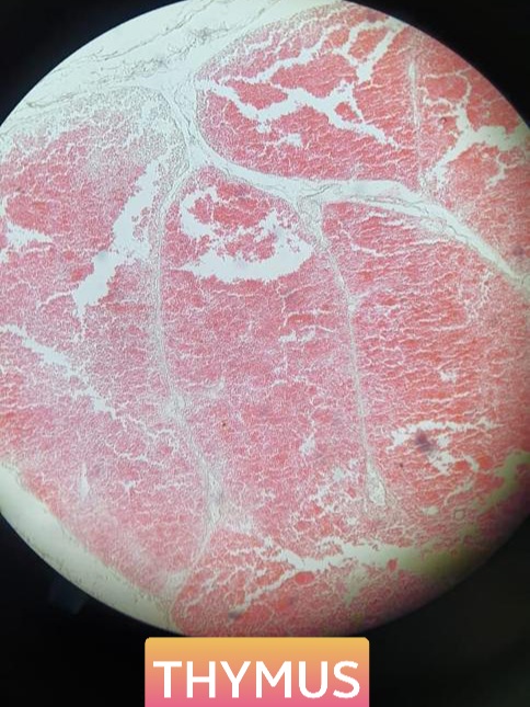
Points of identification:
- Presence of lobules with lymphoid tissue.
- Presence of Hassall’s corpuscles in the medulla.
Spleen

Points of identification:
- Presence of thick capsule and trabeculae.
- Red pulp containing splenic cords and sinusoids and white pulp present.
Palatine tonsil

Points of identification:
- Presence of crypts.
- Presence of subepithelial lymphoid nodule.
Integumentary System
Thin skin

Points of identification:
- Presence of dermis and epidermis.
- Presence of dermis with hair follicles and sebaceous glands.
Thick skin

Points of identification:
- Presence of dermis and thick epidermis.
- Presence of dermis with sweat glands.
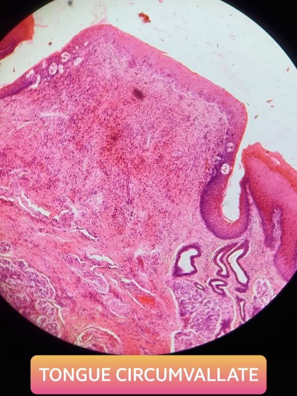
Tongue
Points of identification:
- Presence of different kinds of papillae with skeletal muscle fibers.
- Presence of glands and stratified nonkeratinized squamous cell lining.
Fungiform, filliform papillae

Circumvallate papillae
Endocrine Glands
Pituitary gland
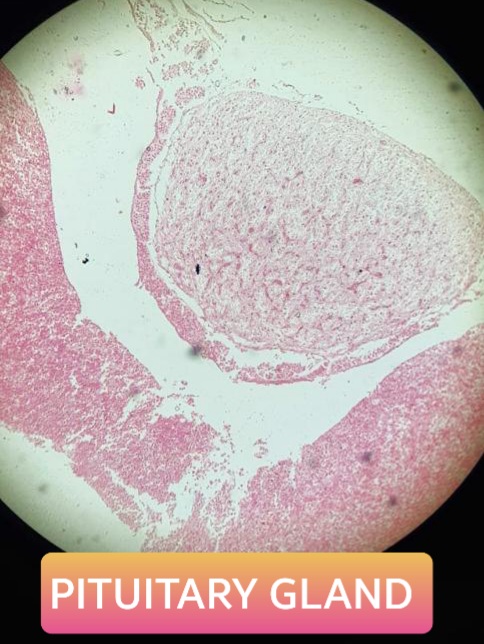
Points of identification:
- Presence of pars anterior with acidophils and basophils.
- Presence of pars nervosa with pituicytes and pars intermedius with colloid.
Thyroid gland
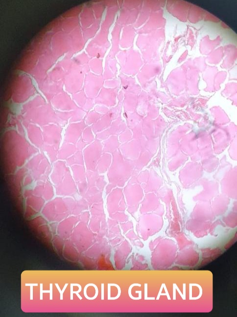
Points of identification:
- Presence of thyroid follicles filled with colloid.
- Presence of parafollicular cells.
Parathyroid gland

Points of identification:
- Cords of cells with numerous sinusoids.
- 2 types of cells – chief (smaller, more in number, central large spherical nuclei) and oxyntic cells (larger, lesser in number).
Suprarenal gland
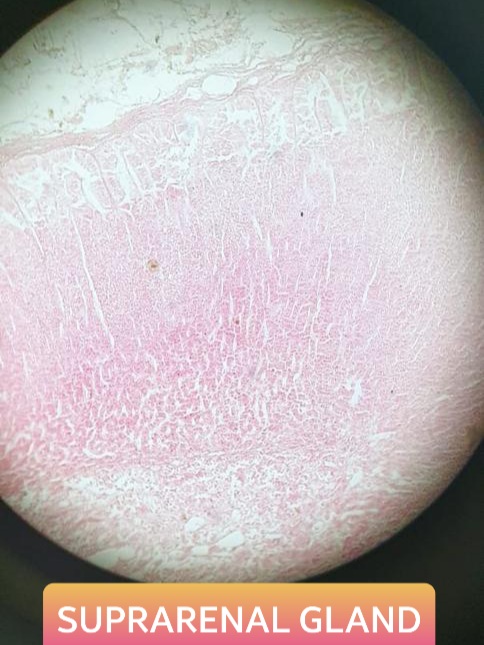
Points of identification:
- Presence of cortex and medulla.
- Presence of secretory cells and sympathetic neurons.
Special Senses
Cornea
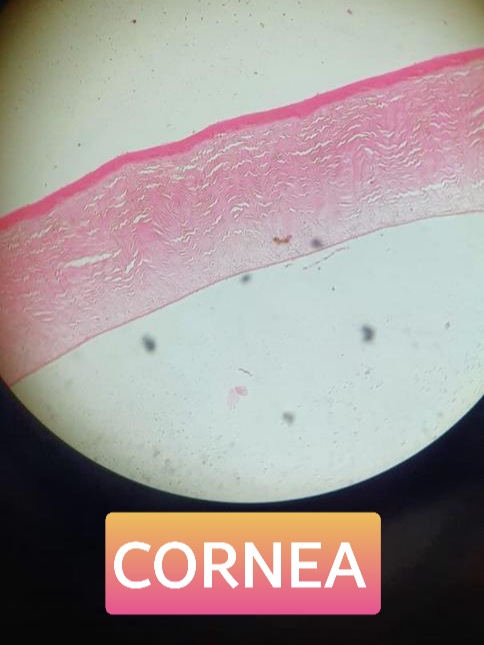
Points of identification:
- Presence of anterior limiting lamina.
- Presence of thick substantia propria with keratocytes.
Retina

Points of identification:
- Presence of 10 layers.
- Presence of external and internal limiting lamina.
Central Nervous System
Spinal cord

Points of identification:
- Presence of H shaped gray mater.
- Presence of central canal.
Cerebellum

Points of identification:
- Presence of molecular layer and granular layer and white mater.
- Presence of Purkinje cells in Purkinje cell layer.
Cerebrum

Points of identification:
- Presence of superficial pia mater and inner white mater.
- Presence of stellate cells and giant pyramidal cells.
Respiratory System
Epiglottis

Points of identification:
- Yellow elastic cartilage present.
- Yellow elastic cartilage covered by stratified squamous epithelium on the oral side and pseudostratified columnar epithelium on the respiratory side.
Trachea

Points of identification:
- Presence of hyaline cartilaginous plates.
- Presence of mucous and serous glands in lamina propria.
Lung
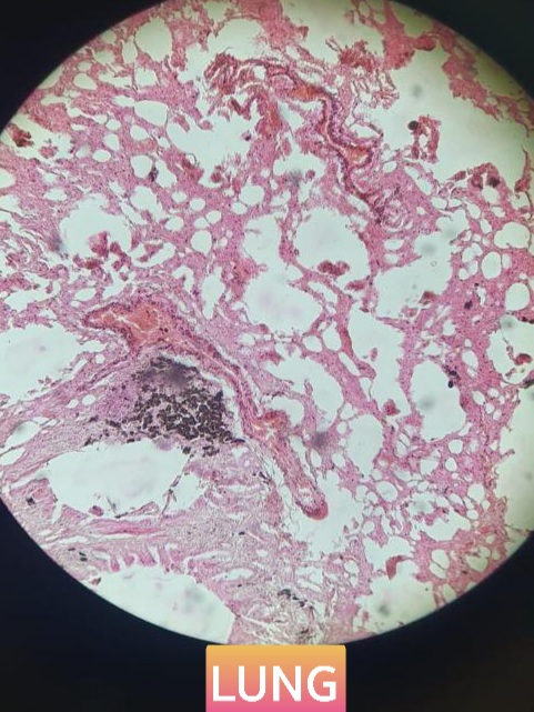
Points of identification:
- Presence of various bronchioles.
- Presence of alveoli lined by simple squamous epithelium.
Gastrointestinal Tract
Esophagus

Points of identification:
- Presence of 4 layers of GIT.
- Presence of esophageal glands in submucosa.
Stomach (fundus)

Points of identification:
- Presence of 4 layers of GIT.
- Presence of fundic glands, gastric pits and mucosal folds.
Stomach (pylorus)

Points of identification:
- Presence of four layers of GIT.
- Presence of pyloric glands, and abundant mucous neck cells.
Duodenum

Points of identification:
- Presence of 4 layers of GIT.
- Presence of Brunner’s glands in submucosa and villi on mucous membrane.
Jejunum

Points of identification:
- Presence of 4 layers of GIT.
- Presence of intestinal crypts and tongue shaped villi.
Ileum

Points of identification:
- Presence of 4 layers of GIT.
- Presence of Peyer’s patches and finger shaped villi.
Large intestine
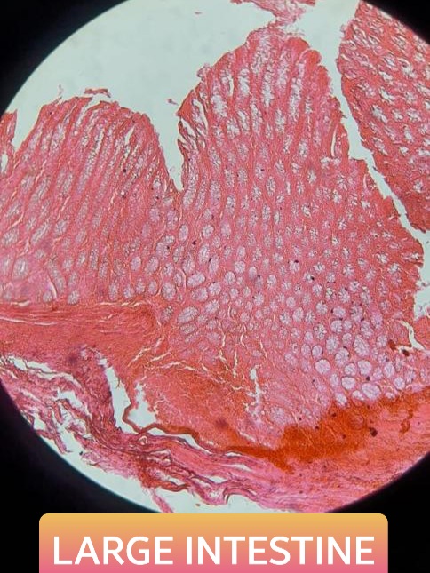
Points of identification:
- Presence of 4 layers of GIT.
- Presence of taenia coli and tubular glands in folds with goblet cells.
Appendix
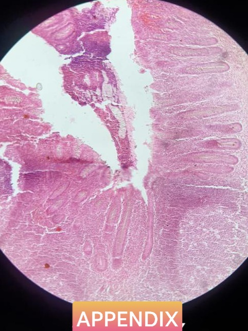
Points of identification:
- Presence of 4 layers of GIT.
- Lymphatic nodules present in submucosa.
Liver

Points of identification:
- Hexagonally arranged hepatocytes with portal triad.
- Presence of central vein.
Gallbladder

Points of identification:
- Presence of 3 layers of GIT.
- Absence of submucosa.
Pancreas

Points of identification:
- Presence of pancreatic acini.
- Presence of islets of Langerhans.
Urinary System
Kidney
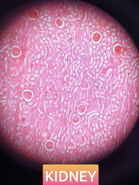
Points of identification:
- Presence of cortex and medulla with cut sections of PCT, DCT, etc.
- Presence of medullary rays and renal corpuscles.
Ureter

Points of identification:
- Presence of star shaped lumen lined by transitional epithelium.
- Presence of 3 muscle coats.
Urinary bladder

Points of identification:
- Presence of transitional epithelial lining.
- Presence of ill-defined muscle coat
Male Reproductive System
Testis

Points of identification:
- Presence of seminiferous tubules with spermatozoa.
- Presence of interstitial cells between the tubules.
Epididymis

Points of identification:
- Presence of highly convoluted efferent ductules with stereocilia.
- Presence of smooth muscle around the ductules.
Vas deferens

Points of identification:
- Presence of narrow irregular lumen with mucosal folds.
- Presence of thick circularly arranged muscle coat.
Prostate

Points of identification:
- Presence of prostatic acini separated by fibromuscular tissue.
- Presence of amyloid body
Female Reproductive System
Ovary
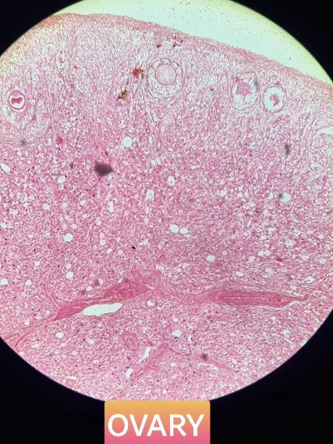
Points of identification:
- Presence of follicles with oocytes in diff erent stages of maturity.
- Presence of germinal epithelium
Uterine tube

Points of identification:
- Presence of primary, secondary, tertiary mucosal folds and its lumen.
- Presence of circular muscle coat.
Uterus
Points of identification:
- Presence of thick myometrium with smooth muscle.
- Presence of uterine glands in endometrium.
Placenta

Points of identification:
- Chorionic villi of different stages seen.
- Intervillous space filled with maternal blood and RBCs.
Umbilical cord
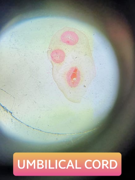
Points of identification:
- Presence of 2 umbilical arteries and 1 umbilical vein.
- Presence of Wharton’s jelly.
Hope you found the article helpful! Do share it with your batchmates.
Good luck Medicoholics! Until next time.
Thank you so much. I need tips, question papers for all subjects of 1st year mbbs.
You’re welcome!
For more such useful articles, click on the MBBS option on the top menu.
Pdf plz
You can download the pdf.
Thanks a lot😊
Thank you. It was a life saver blog
Very helpful. How can I download this?
By clicking on the “download pdf” button at the bottom of the page.